2-Year Expanded Efficacy Data From the GATHER2 Trial:
Advances in Dry AMD:
“2-Year Expanded Efficacy Data From the GATHER2 Trial” The GATHER2 trial provided two-year expanded efficacy data on the investigational treatment for geographic atrophy secondary to age-related macular degeneration. The treatment involves avacincaptad pegol (ACP), which is an inhibitor of the C5 protein in the immune system. The results demonstrated sustained improvements in visual function and a significant reduction in lesion growth compared to the control group. Over the two years, patients receiving the treatment exhibited a meaningful preservation of vision, with a favorable safety profile. These findings underscore the potential of the therapy to provide long-term benefits for individuals affected by this progressive retinal disease.
Khanani AM, Patel SS, Staurenghi G, Tadayoni R, Danzig CJ, Eichenbaum DA, Hsu J, Wykoff CC, Heier JS, Lally DR, Monés J, Nielsen JS, Sheth VS, Kaiser PK, Clark J, Zhu L, Patel H, Tang J, Desai D, Jaffe GJ; GATHER2 trial investigators. Efficacy and safety of avacincaptad pegol in patients with geographic atrophy (GATHER2): 12-month results from a randomised, double-masked, phase 3 trial. Lancet. 2023 Oct 21;402(10411):1449-1458. doi: 10.1016/S0140-6736(23)01583-0. Epub 2023 Sep 8. PMID: 37696275.
This case report describes a 42-year-old male who was referred by an optometrist to our ophthalmologist Dr. Shah with an asymptomatic left inferotemporal cystic lesion with vascular bulbs at the tips, discovered during a routine eye examination. Differential diagnosis includes cavernous hemangioma associated with intracranial vascular anomalies, and retinal astrocytic hamartoma associated with tuberous sclerosis. The importance of ruling out associated intracranial lesions, including vascular malformations and tubers, is emphasized.
Introduction
The patient, a 42-year-old Caucasian male, was referred for a detailed ophthalmological evaluation after a cystic lesion was identified during a routine eye exam. The patient had no complaints of vision loss, floaters, or any other symptoms. There was no significant past medical history and no known family history of eye diseases or systemic conditions like tuberous sclerosis.
Clinical Findings
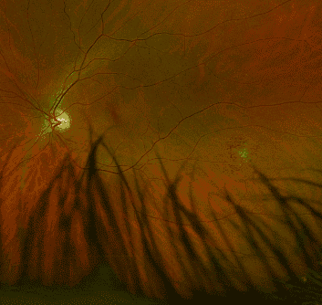
Upon examination, visual acuity was 20/20 in both eyes, and intraocular pressure was within normal limits. Slit-lamp examination was unremarkable in both eyes. Dilated fundus examination revealed a lesion in the inferotemporal region of the left eye. The lesion appeared cystic, with distinct vascular bulbs at the tips. No other abnormalities were noted in either eye.
Diagnostic Tests
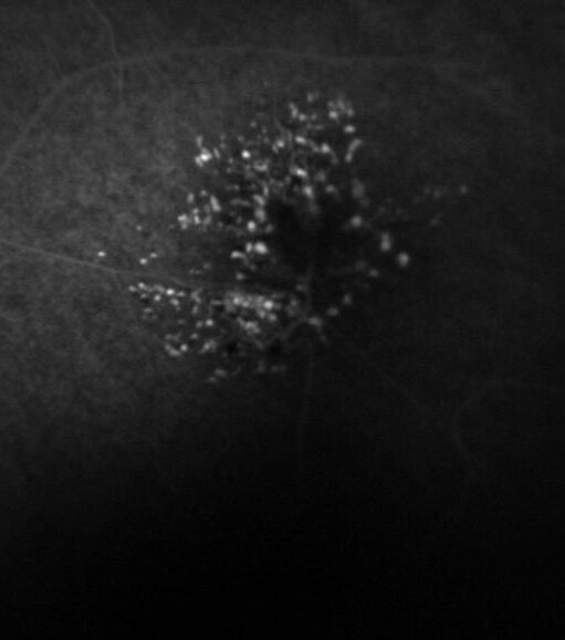
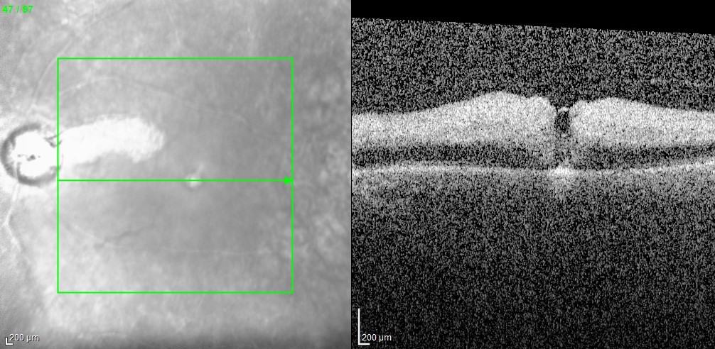
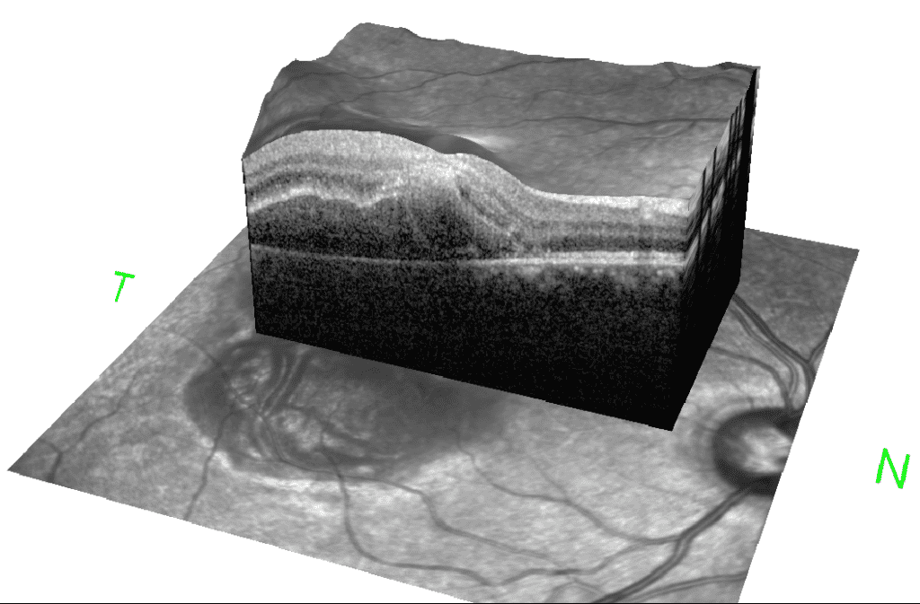
OCT (Optical Coherence Tomography) confirmed the central cystic nature of the lesion with surrounding retinal tissue appearing structurally normal. Fluorescein angiography showed hyperfluorescence with late staining, but no leakage. MRI of the orbit and brain was recommended to rule out intracranial involvement.
Differential Diagnosis
Cavernous Hemangioma associated with Intracranial Vascular Anomalies: The cystic nature of the lesion with vascular bulbs may be indicative of a cavernous hemangioma. These are known to occur sporadically but may also be associated with intracranial vascular malformations.
Retinal Astrocytic Hamartoma associated with Tuberous Sclerosis: Given the patient’s age and the appearance of the lesion, retinal astrocytic hamartoma was also considered. These benign lesions are often associated with tuberous sclerosis and can have similar clinical features.
Retinal Vaso-proliferative tumors associated with sub-retinal exudation and potential subretinal fibrosis. These tumors can be distinguished from Cavernous Hemangioma by the absence of vascular bulbs, and from Retinal Astrocytic Hamartoma by not showing the characteristic calcified retinal nodules associated with Tuberous Sclerosis.
Importance of Ruling out Intracranial Lesions
The presence of either a cavernous hemangioma or an astrocytic hamartoma raises the suspicion for associated intracranial lesions such as vascular malformations in the former and tubers in the latter. Failure to identify these intracranial anomalies may lead to severe neurologic consequences, including seizures, hemorrhages, or more serious complications.
Management and Follow-up
Given the uncertainty of the diagnosis, the patient was referred for an MRI of the orbit and brain, and consultation with a neurologist was recommended for further evaluation of potential intracranial lesions. Genetic testing for markers of tuberous sclerosis was also advised.
Conclusion
This case emphasizes the significance of comprehensive evaluation for patients presenting with retinal lesions. A multidisciplinary approach involving retina specialists, other eye doctors, and neurologists. This can be crucial for accurate diagnosis and management, especially in cases where intracranial involvement is suspected.
References
Shields JA, Shields CL. Atlas of Intraocular Tumors. Lippincott Williams & Wilkins; 1999.
Introduction: Presented in this case is a male patient born in 1952 who was initially admitted for a stroke on August 1st. The patient’s new chief complaint upon re-admission was decreased vision in the left eye since August 17th. This report focuses on the ophthalmological evaluation and management of the patient’s left eye vision loss.
Case Presentation: The patient, a 71-year-old male, presented to our facility with a history of stroke on August 1st. He complained of decreased vision in his left eye since August 17th. He was re-admitted to our hospital for further evaluation. Due to the concerning visual symptoms, an ophthalmologist was consulted for a comprehensive eye examination.
Ophthalmological Examination: Upon examination by our ophthalmologist, Dr. Shah, the patient’s visual acuity in the left eye was found to be reduced to 20/40. Fundoscopic examination revealed a left eye cilioretinal artery perfused with a central retinal artery occlusion. Further scrutiny showed the presence of several cholesterol plaques in the retinal arterioles, suggestive of an embolic event. Optical Coherence Tomography (OCT) scan of the left retina showed inner retinal edema with preservation of the outer retina, particularly in the distribution of the cilioretinal artery. Additionally, infrared imaging demonstrated retinal pigment epithelium preservation corresponding to the distribution of the cilioretinal artery.
Discussion: The patient’s medical history included a previous stroke for which he underwent thrombectomy. Unfortunately, the thrombectomy resulted in a subarachnoid hemorrhage due to reperfusion, rendering double anticoagulation contraindicated. Despite the occurrence of a new embolic event leading to a central retinal artery occlusion, the primary concern remained the patient’s overall life. Therefore, the decision was made to postpone double anticoagulation until the subarachnoid hemorrhage had resolved. The ophthalmologist emphasized that, in such cases, life must take precedence over vision.
Conservative Measures and Management Options: Given the constraints of anticoagulation, the patient’s management included conservative measures to attempt reperfusion of the retina. These measures were tailored to the patient’s unique medical history and risk profile. The rarity of the situation was underscored by the ophthalmologist, who highlighted the fortunate preservation of central vision thanks to the rare presence of the cilioretinal artery, which only 15% of patients have. Normally, vision is decreased to 20/200 or worse in a central retinal artery occlusion without a cilioretinal artery present. The patient’s intraocular pressure will be reduced with topical drops to maintain perfusion to the fovea and remaining retina. The patient will also be monitored for neovascularization of the retina and angle to prevent “90 day glaucoma.”
Conclusion: In conclusion, this case underscores the intricate interplay between ophthalmological concerns and the patient’s overall medical condition. Central retinal artery occlusions can present in the context of complex medical histories, requiring tailored management approaches. The rarity of a perfused cilioretinal artery, which played a critical role in preserving central vision, highlights the importance of early ophthalmological evaluation and collaboration with specialists to optimize patient outcomes.
Patient Details: A 32-year-old male presented to our ophthalmologist’s retina clinic with a history of a traumatic injury to his right eye. The patient reported being hit in the right eye with a beer bottle, resulting in immediate visual impairment and pain.
Clinical Presentation: Upon examination, the patient exhibited reduced visual acuity in the right eye, with a visual acuity of Count Finger (CF) at one foot. Slit-lamp biomicroscopy revealed a localized foveo-macular retinal detachment with a subretinal hemorrhage, consistent with a choroidal rupture. Fundus examination confirmed the presence of a choroidal rupture associated with the macular region. No signs of proliferative vitreoretinopathy or other retinal pathologies were noted in the fellow eye.
Treatment Approach: Dr. Shah, a retinal ophthalmologist, was consulted for the management of this challenging case. After a thorough discussion of treatment options and risks, a decision was made to proceed with a pneumatic retinopexy. The procedure involved the injection of 0.3 cc’s of perfluoropropane (C3F8) gas into the vitreous cavity, followed by face-down positioning to promote retinal reattachment.
Outcome: Following the pneumatic retinopexy and adherence to the prescribed face-down positioning regimen, the patient exhibited remarkable improvement in his visual acuity. Serial examinations demonstrated successful reattachment of the fovea, with visual acuity improving from Count Finger at one foot to 20/40. Despite mild perifoveal scarring, the patient’s visual function showed a significant and favorable outcome.
Discussion: Choroidal ruptures are a rare consequence of blunt ocular trauma and can lead to various retinal complications, including retinal detachment. In this case, the localized foveo-macular retinal detachment resulting from a choroidal rupture required a tailored intervention to promote retinal reattachment. Pneumatic retinopexy with the administration of C3F8 gas, combined with face-down positioning, proved to be an effective strategy in achieving successful anatomical and functional outcomes.
It is important to note that despite the successful reattachment, mild perifoveal scarring was observed. While this scarring may contribute to some residual visual impairment, the overall improvement in visual acuity signifies a significant therapeutic success.
Future Considerations: The patient’s clinical course warrants ongoing monitoring, as a new choroidal neovascular membrane has been identified. Given the potential for further visual impairment and complications, the patient may benefit from anti-vascular endothelial growth factor (anti-VEGF) therapy by a retinal ophthalmologist. This targeted treatment approach aims to inhibit neovascularization and preserve visual function in the presence of choroidal neovascularization.
In conclusion, the presented case highlights the successful management of a foveo-macular retinal detachment secondary to a choroidal rupture through pneumatic retinopexy and face-down positioning. The patient’s substantial improvement in visual acuity, despite mild scarring, underscores the importance of tailored therapeutic interventions in cases of traumatic ocular injury involving choroidal breaks. Continued vigilance and appropriate intervention, such as anti-VEGF therapy, will be essential in ensuring optimal visual outcomes for the patient.
Faricimab is a treatment for macular degeneration, specifically targeting Angiopoietins and the Tie-2 Pathway. Here we discuss a review of relevant clinical trials led by Dr. Arshad Khanani.
The review article PDF is available for download here, and below is a concise summary as well as additional, helpful resources related to Faricimab.
Angiopoietins and Tie-2 Pathway: Initially, the review discusses angiopoietins (Ang-1 and Ang-2) and their interactions with the Tie-2 receptor. Ang-2 acts as a partial agonist or antagonist, and its inhibition consequently reduces vascular leakage and improves organ function.
Vascular and Extravascular Functions of Angiopoietin-2: In addition to the above, Ang-2 has functions in both vascular and extravascular settings for macular degeneration. Particularly in hyperglycemia and hypoxia, it plays a role in stimulating pericyte loss, neovascularization, and the breakdown of the Blood Retinal Barrier (BRB).
Diabetic Retinopathy and Macular Edema: Moreover, the article explores Ang-2’s role in macular degeneration, diabetic retinopathy and diabetic macular edema (DME). It emphasizes the importance of the retinal vasculature and the sensitivity of the retina to pericyte loss.
Angiopoietin-Like Proteins (ANGPTL) and Metabolism: Additionally, the paper delves into ANGPTL, which are similar to angiopoietins but have distinct roles in glucose and lipid metabolism. Specifically, ANGPTL-3, ANGPTL-4, and ANGPTL-8 are discussed in this context.
Treatment and Therapeutic Targets: The document also mentions various treatments and innovative therapies for neovascular age-related macular degeneration and diabetic retinopathy, including surgical innovations.
Review:
The systematic review article provides a comprehensive analysis of angiopoietins and their role in macular degeneration, vascular biology, inflammation, and metabolic processes. Notably, the focus on diabetic retinopathy and the connection between angiopoietins and metabolic disorders adds significant value.
The paper’s strength lies in its integration of various studies and research findings, thereby providing a holistic view. Furthermore, the exploration of ANGPTL and their role in metabolic processes is particularly intriguing and opens avenues for further research.
However, the study might be challenging for a lay reader due to its technical language and complex concepts. Therefore, its target audiences are medical professionals or researchers in the field of biology, medicine, or related disciplines.
Overall, this review article is a valuable resource for those interested in the intricate interplay between angiopoietins, vascular biology, and metabolic disorders. Consequently, it offers insights that could potentially lead to new therapeutic approaches for conditions like diabetic retinopathy and metabolic syndromes.
A 54-year-old female patient visited the clinic for the evaluation of potential macular degeneration with choroidal neovascular membrane in her left eye. The referring physician suggested injecting anti-VEGF into the left eye to treat “wet” macular degeneration. For a few months, the patient had been experiencing intermittent blurry vision in her left eye associated headaches, and floaters. The left eye had a mild afferent pupillary defect and extensive pigmentary atrophy centrally. During the examination, subretinal pigmentary changes and drusen were present in both maculae. Fundus photos are shown below:
Figure 1: Fundus montages imaging in both eyes. Posterior pigmentary changes are visible in both eyes, with drastically more changes in the left eye. Also, the left eye displays peripheral white-yellow lesions.
To better understand the disease, fluorescein angiogram, fundus autofluorescence, and indocyanine green angiography were performed. These were done with regards to the left eye’s asymmetry and peripheral lesions.
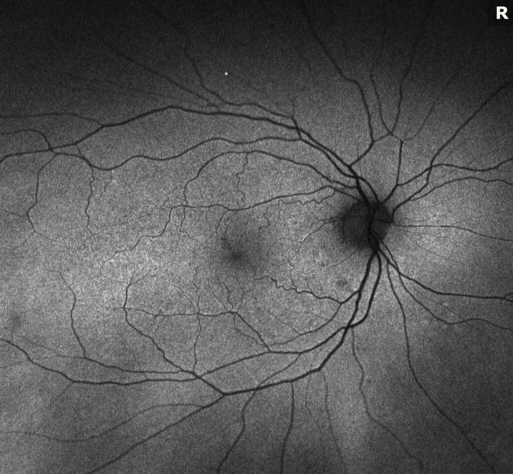
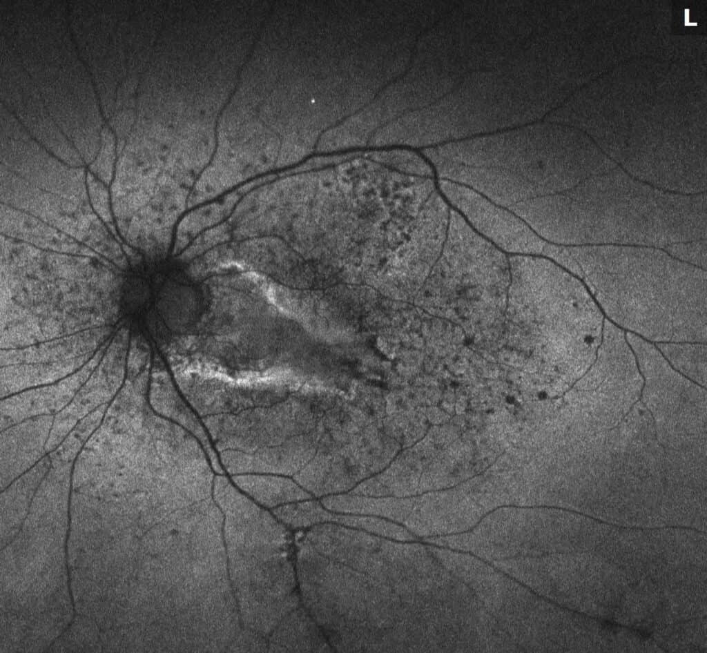
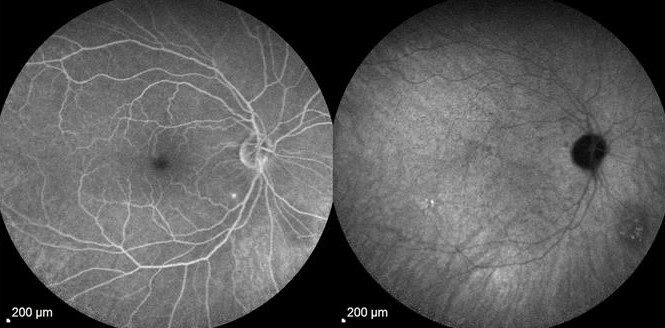
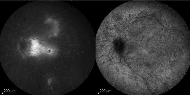
Figure 2: Fundus autofluorescence images. The right eye displays normal autofluorescence consistent with drusen. The left eye shows mixed hyper- and hypo-autofluorescence, which are seen with both subretinal fibrosis and drusen.
Figure 3: Fluorescein angiogram (FA) with indocyanine green angiography (ICG) images. The right eye displays late staining of drusen, which is visible on FA. There is a hypocyanescence inferonasal to the nerve which is seen with a choroidal lesion of unknown significance on ICG. Early staining of subretinal fibrosis and late leakage of cystoid macular edema are visible.
Additional history was obtained from the patient about peripheral lesions, asymmetry of disease, and extensive scarring of the left eye in a pattern that is not normal for AMD. One year ago, the patient reported sudden loss of vision and was referred to a neuro-ophthalmologist with the possibility of optic neuritis or ischemic optic neuropathy. She was treated with IV Solumedrol for 3 days with improvement but then did not make it to a follow-up appointment and realized drastic vision loss. The retinal lesions were taken note of during her next visit to her comprehensive ophthalmologist, who then referred her to our office.
The afferent pupillary defect indicated a history of optic nerve inflammation. The differential diagnosis was broadened from the referral for choroidal neovascular membrane related to macular degeneration due to the white dots in the periphery and subretinal fibrosis. Additional lab testing aided in narrowing down to non-infectious entities. Oral steroids, the typical treatment in MFCPU, were begun. In order to diminish her cystoid macular edema, local anti-inflammatory and anti-VEGF injections were used. The cystoid macular edema improved after this. Unfortunately, the subretinal fibrosis in the macula slowly stopped progressing. The patient was given steroid sparing immunosuppressive therapy (methotrexate) in response, and she is doing well.
MFCPU is an idiopathic, inflammatory disease that results in bilateral inflammation in an episodic pattern. Patients with this disease typically have multiple chorioretinal lesions (unseen in our patient). The white dots in this disease are much bigger than those that are visible in punctate inner choroidopathy (PIC). In opposition to presumed ocular histoplasmosis syndrome (POHS), vitritis is common. Treatment can help avoid full vision loss. The general eye professionals were right in seeing and identifying drusen in her eyes, but the patient also had a more subtle, uveitic reason for her left retinal fluid, which could have been treated earlier at our clinic to prevent additional fibrosis/scarring. Early referral to a retina specialist is vital, as seen in this case.
References
1. Essex, Rohan W., et al. “Idiopathic Multifocal Choroiditis.” Retina, vol. 33, no. 1, 2013, pp. 1–4., doi:10.1097/iae.0b013e3182641860.
2. Goldstein, Debra, and Lary Ulanski. “Multifocal Choroiditis vs. PIC: Variations on a Theme?” Review of Ophtalmology, 2004.
3. Pinar, Vakur, and Stephen Foster. “Bilateral Chorioretinal Lesions Appearing Late in Posterior Uveitis.” 2000.
4. Dolz-Marco, Rosa, et al. “How to Differentiate Myopic Choroidal Neovascularization, Idiopathic Multifocal Choroiditis, and Punctate Inner Choroidopathy Using Clinical and Multimodal Imaging Findings.” Ophthalmic Surgery, Lasers and Imaging Retina, SLACK Incorporated, 16 Mar. 2017,
Macular degeneration is a prevalent eye condition that affects millions of people worldwide, leading to vision loss and decreased quality of life. In the quest for more effective treatments, medical research has made significant strides in developing new medications to combat this debilitating disease. At the forefront of these advancements is Dr. Shah’s clinic, where cutting-edge treatments for macular degeneration are being offered. Dr. Shah is not only a leading expert in the field but also actively involved in clinical trials, bringing hope and potential relief to countless patients.
The Need for New Medications
Macular degeneration is typically divided into two main types: dry and wet. The dry form, while more common, lacks definitive treatment options. This has driven researchers and medical professionals to focus their efforts on the wet form, characterized by abnormal blood vessel growth in the retina. Conventional treatment for wet macular degeneration involves regular intravitreal injections of anti-VEGF medications. However, not all patients respond equally to this treatment, and its frequent administration can be burdensome.
New Medications on the Horizon
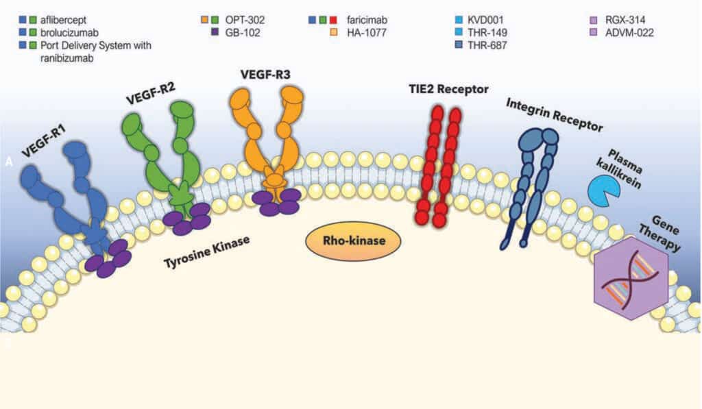
Targeted Complement Inhibitors: Researchers have identified that dysregulation of the complement system plays a significant role in wet AMD development. By inhibiting specific components of this system, targeted complement inhibitors offer a novel approach to managing the disease. These medications have shown promising results in clinical trials, demonstrating their potential to provide more personalized and effective treatment for patients.
Tyrosine Kinase (TK) Inhibitors: TK is another protein involved in the abnormal blood vessel growth seen in wet AMD. TK inhibitors have emerged as potential adjunctive therapies to anti-VEGF medications. Clinical trials have shown that combining TK inhibitors with anti-VEGF treatments can lead to improved outcomes, potentially reducing the frequency of injections and enhancing patient satisfaction.
Dr. Shah’s Contributions to Advancements
As a distinguished ophthalmologist specializing in retinal diseases, Dr. Shah has been at the forefront of introducing new medications for macular degeneration in his clinic. His commitment to staying current with the latest research and advancements has led him to actively participate in clinical trials. By participating in these trials, Dr. Shah is not only offering his patients access to cutting-edge treatments but also contributing to the advancement of medical knowledge and the potential for better outcomes for all patients with macular degeneration.
The Importance of Clinical Trials
Clinical trials are a crucial aspect of medical research and innovation. Through these trials, new medications undergo rigorous testing to determine their safety and effectiveness. By being part of clinical trials, Dr. Shah is playing a vital role in shaping the future of macular degeneration treatment. Patients who choose to participate in these trials not only have access to potential breakthrough therapies but also contribute to the collective knowledge that will ultimately benefit countless others facing macular degeneration.
Conclusion
Macular degeneration is a sight-threatening condition that demands constant exploration and innovation in treatment options. With new medications on the horizon, patients have reason to be hopeful. Dr. Shah’s clinic stands at the forefront of these developments, offering cutting-edge treatments and actively participating in clinical trials to bring advancements from the research lab to the patient’s bedside. As we look forward to a brighter future for macular degeneration patients, it is reassuring to know that dedicated professionals like Dr. Shah are working tirelessly to improve outcomes and quality of life for those affected by this challenging condition. If you or a loved one is struggling with macular degeneration, consider consulting with Dr. Shah to explore the latest treatment options and potential participation in clinical trials for a chance at a better tomorrow.